MMM
YYYY
3D imaging lipidometry in single cell by in-flow holographic tomography
流内全息层析成像在单细胞中的三维成像测脂
単細胞中の流内ホログラフィートモグラフィーによる3次元イメージング脂肪測定
유내 홀로그램 계층 분석 영상이 단세포 중의 3차원 영상 지방 측정
Medición de la grasa mediante tomografía holográfica de flujo en una sola célula
Imagerie tridimensionnelle des lipides par tomographie holographique en flux dans une seule cellule
голографический анализ изображений в одной клетке
Daniele Pirone ¹ ², Daniele Sirico ¹ ³, Lisa Miccio ¹, Vittorio Bianco ¹, Martina Mugnano ¹, Danila del Giudice ¹ ⁴, Gianandrea Pasquinelli ⁵ ⁶, Sabrina Valente ⁵, Silvia Lemma ⁷ ⁸ ⁹, Luisa Iommarini ⁹ ¹⁰, Ivana Kurelac ⁷ ⁸ ⁹, Pasquale Memmolo ¹, Pietro Ferraro ¹
¹ CNR-ISASI, Institute of Applied Sciences and Intelligent Systems “E. Caianiello”, Via Campi Flegrei 34, Pozzuoli, Napoli 80078, Italy
² Department of Electrical Engineering and Information Technologies, University of Naples “Federico II”, via Claudio 21, Napoli 80125, Italy
³ Department of Chemical, Materials and Production Engineering of the University of Naples Federico II, Piazzale Tecchio 80, Napoli 80125, Italy
⁴ Department of Mathematics and Physics, University of Campania “Luigi Vanvitelli”, Caserta 81100, Italy
⁵ Biotechnology and Methods in Laboratory Medicine, Department of Experimental, Diagnostic and Specialty Medicine (DIMES), University of Bologna, Bologna 40126, Italy
⁶ Subcellular Nephro-Vascular Diagnostic Program, Pathology Unit S.Orsola IRCCS, University of Bologna, Bologna 40126, Italy
⁷ Unit of Medical Genetics, Department of Medical and Surgical Sciences (DIMEC), University of Bologna, Via Massarenti 9, Bologna 40138, Italy
⁸ Study and Research Center on Gynecological Neoplasias, Department of Medical and Surgical Sciences (DIMEC), University of Bologna, Via Massarenti 9, Bologna 40138, Italy
⁹ Center for Applied Biomedical Research (CRBA), University of Bologna, Bologna 40138, Italy
¹⁰ Department of Pharmacy and Biotechnology (FABIT), University of Bologna, Bologna 40126, Italy
Opto-Electronic Advances, 25 August 2022
Abstract

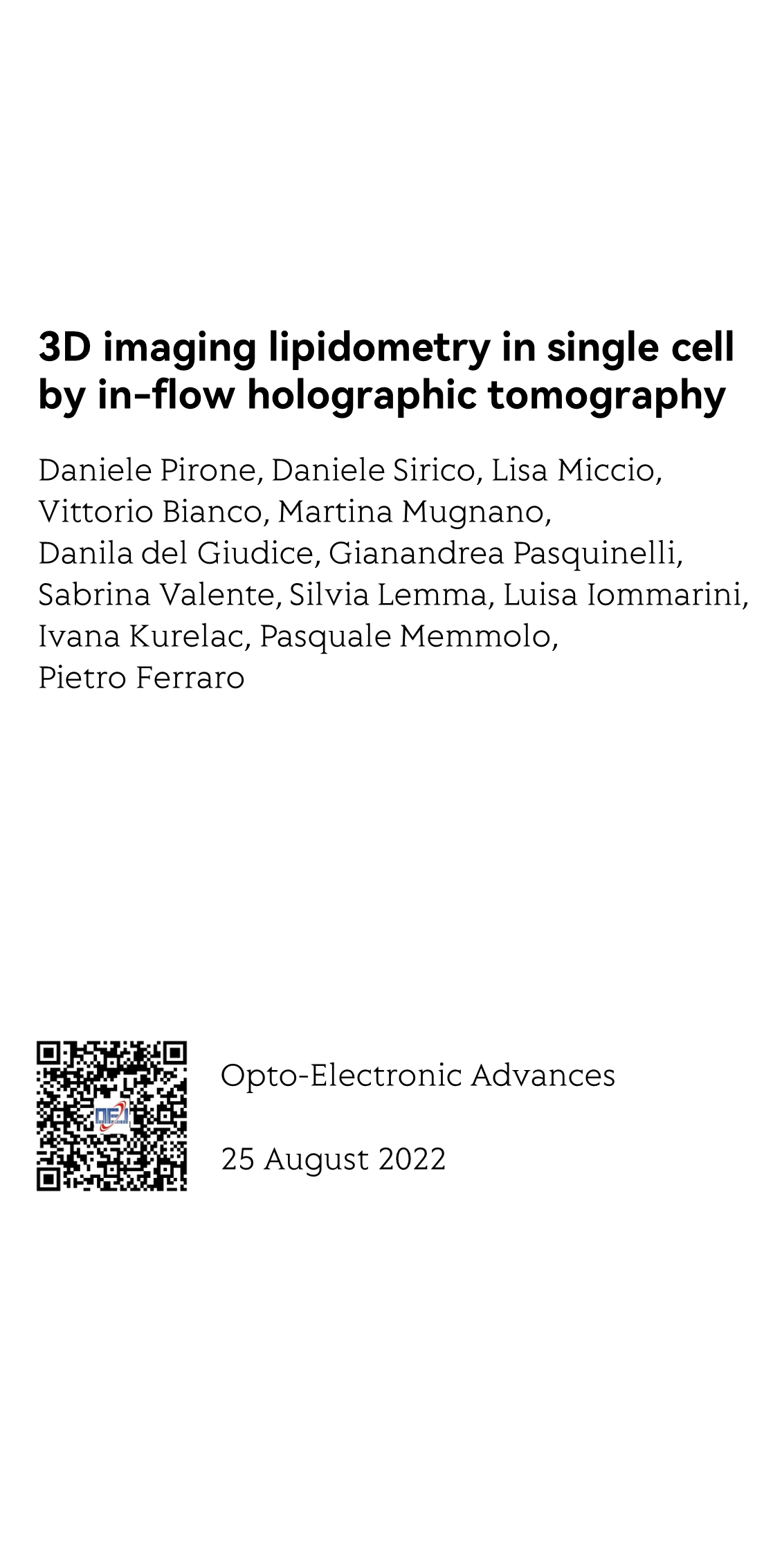
The most recent discoveries in the biochemical field are highlighting the increasingly important role of lipid droplets (LDs) in several regulatory mechanisms in living cells. LDs are dynamic organelles and therefore their complete characterization in terms of number, size, spatial positioning and relative distribution in the cell volume can shed light on the roles played by LDs.

Until now, fluorescence microscopy and transmission electron microscopy are assessed as the gold standard methods for identifying LDs due to their high sensitivity and specificity. However, such methods generally only provide 2D assays and partial measurements. Furthermore, both can be destructive and with low productivity, thus limiting analysis of large cell numbers in a sample.

Here we demonstrate for the first time the capability of 3D visualization and the full LD characterization in high-throughput with a tomographic phase-contrast flow-cytometer, by using ovarian cancer cells and monocyte cell lines as models. A strategy for retrieving significant parameters on spatial correlations and LD 3D positioning inside each cell volume is reported.

The information gathered by this new method could allow more in depth understanding and lead to new discoveries on how LDs are correlated to cellular functions.
Opto-Electronic Advances_1
Opto-Electronic Advances_2
Opto-Electronic Advances_3
Opto-Electronic Advances_4
Reviews and Discussions
https://www.hotpaper.io/index.html
High-intensity spatial-mode steerable frequency up-converter toward on-chip integration
Unraveling the efficiency losses and improving methods in quantum dot-based infrared up-conversion photodetectors
Ultrafast dynamics of femtosecond laser-induced high spatial frequency periodic structures on silicon surfaces
Optical scanning endoscope via a single multimode optical fiber
Simultaneously realizing thermal and electromagnetic cloaking by multi-physical null medium
Data-driven polarimetric imaging: a review
Robust measurement of orbital angular momentum of a partially coherent vortex beam under amplitude and phase perturbations
Deblurring, artifact-free optical coherence tomography with deconvolution-random phase modulation
Inverse design for material anisotropy and its application for a compact X-cut TFLN on-chip wavelength demultiplexer
Improved spatiotemporal resolution of anti-scattering super-resolution label-free microscopy via synthetic wave 3D metalens imaging
Flat soliton microcomb source
Smart palm-size optofluidic hematology analyzer for automated imaging-based leukocyte concentration detection


Previous Article                                Next Article
About
|
Contact
|
Copyright © Hot Paper